Successfully Added to Cart
Updated Cart Total:
0
items
Subtotal:
$0.00 USD
Customers also bought...

-
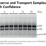 DNA/RNA Shield (50 ml)Cat#: R1100-50DNA/RNA Shield reagent is a DNA and RNA stabilization solution for nucleic acids in any biological sample. This DNA and RNA stabilization solution preserves the...
 -
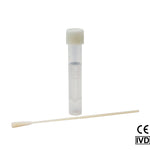 DNA/RNA Shield SafeCollect Swab Collection Kit (1 ml fill) (1 collection kit)Cat#: R1160The DNA/RNA Shield SafeCollect Swab Collection Kit is a user-friendly collection kit for stabilizing the nucleic acid content of samples collected with a swab. DNA/RNA...


Highlights
- 100 T Equivalent: Prepared from Arthrobacter lutes. Essential enzyme activities are β-1,3-glucanase and β-1,3-glucan laminaripentao-hydrolase.
- Convenient: Provided lyophilized along with a storage buffer for reconstitution.
- Efficient Cell Wall Digestion: Supplied storage buffer has been optimized to confer maximum levels of enzymatic activity.


Original Manufacturer
100% satisfaction guaranteed, read Our Promise
Innovated in California, Made in the USA
Highlights
- 100 T Equivalent: Prepared from Arthrobacter lutes. Essential enzyme activities are β-1,3-glucanase and β-1,3-glucan laminaripentao-hydrolase.
- Convenient: Provided lyophilized along with a storage buffer for reconstitution.
- Efficient Cell Wall Digestion: Supplied storage buffer has been optimized to confer maximum levels of enzymatic activity.

Original Manufacturer
100% satisfaction guaranteed, read Our Promise
Innovated in California, Made in the USA
Description
Performance
Technical Specifications
| Activity | β-1,3-glucan laminaripentao-hydrolase and β-1,3-glucanase (trace amount of protease, ca. 1.5 units per 10 µl). DNase and RNase: none detectable. |
|---|---|
| Concentration | 5 U/µl |
| Deactivation | Lytic activity is lost in 5 minutes at 60°C |
| Enzyme Source | Arthrobactor luteus |
| Format | Lyophilized enzyme provided w/ storage buffer |
| Optimum pH & Temp | pH 7.5, 35°C (lysis of viable yeast), pH 6.5, 45°C (hydrolysis of yeast glucan). |
| Product Storage | Zymolyase is stable for over 1 year at -20°C or many years below -70°C. |
| Protein Concentration | ~10-15 mg/ml |
| Susceptible Fungal Genera: | Asbya, Candida, Debaryomyces, Eremothecium, Endomyces, Hansenula, Hanseniaspora, Kloekera, Kluyveromyces, Lipomyces, Metschikowia, Pichia, Pullularia, Saccharomyces, Saccharomyces, Saccharomycopsis, Schizosaccahromyces, Torulopsis |
| Unit Definition | One lytic unit is defined as catalyzing a 10% decrease in optical density at 800 nm (OD800) at 30°C in 30 minutes. |
| Usage | Spheroplast/Protoplast formation; Yeast cell fusion; Yeast transformation; Lysis of fungi |
Resources
Documents
FAQ
Zymolyase is very efficient, but here are some tips:
1. Use fresh and early log phase cells, which are more susceptible to the lytic properties of Zymolyase.
2. Using less cells as input can improve overall lysis efficiency.
3. Zymolyase digestion time can also be increased to 2 – 16 hours.
Citations
Need help? Contact Us